
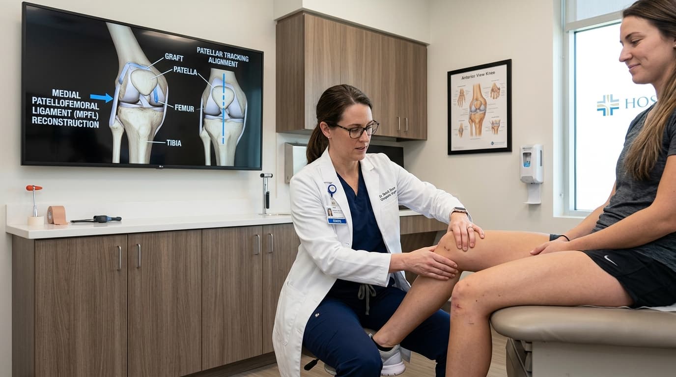

Understanding Patella Instability, Dislocation & MPFL Reconstruction
The patella (kneecap) glides in a specialized groove (trochlea) on the front of the femur. When the main stabilizing restraint—the Medial Patellofemoral Ligament (MPFL)—tears during a twisting knee injury, the kneecap dislocates outwards. Recurrent episodes cause cartilage damage, severe apprehension ('fear of the knee giving out'), and early patellofemoral arthritis. Anatomical MPFL reconstruction replaces the torn ligament with a slender tendon graft, firmly locking the kneecap in its groove throughout motion.
The Minimally Invasive Arthroscopy Advantage
Advanced 4K keyhole arthroscopy allows complete visualization and anatomical repair through pencil-thin incisions, eliminating extensive soft-tissue cutting, preserving natural joint biomechanics, and drastically accelerating recovery.
When Should You Consider Consultation for Patella Instability & MPFL?
Specialist intervention is considered when joint pain or functional limitation significantly disrupts your daily life and conservative measures have been exhausted. Check if you experience any of the following:
First-time or recurrent kneecap dislocation with severe pain, immediate swelling, and visible deformity.
Constant feeling that the kneecap is loose, slipping sideways, or about to pop out during pivoting.
Anterior knee pain, catching, or crunching (crepitus) under the kneecap during stairs or squatting.
Young female athlete or dancer with generalized joint hypermobility and recurrent patellar subluxation.
MRI evidence of MPFL tear, trochlear dysplasia, or osteochondral loose body from the patella.

Dr. Vijay Kumar
Verified SpecialistSenior Consultant Orthopaedic, Robotic Joint Replacement & Complex Articular Trauma Surgeon
“My surgical philosophy centers on biological preservation, anatomical restoration, and rapid functional recovery. By tailoring the intervention to each patient's unique anatomy and lifestyle, we achieve lasting pain relief and active mobility.”
The 4-Stage Precision Clinical Workflow
Pre-Operative MRI & Diagnostic Mapping
4K arthroscopic inspection assessing patellofemoral cartilage congruency and extracting loose bodies.
High-Definition 4K Arthroscopic Access
Harvesting of slender gracilis tendon through a miniature cosmetic incision.
Precision Anatomical Repair / Reconstruction
Anatomical anchor fixation to the upper medial border of the patella.
Bio-Fixation & Sports Rehab Roadmap
Precise isometric femoral tunnel placement at Schottle's point under live fluoroscopy.
Stage 05: Arthroscopic Execution
Controlled graft tensioning ensuring the patella tracks smoothly without over-constraint.
Treatment Modalities Offered
Dr. Vijay Kumar practices conservative joint-sparing protocols first, proceeding to advanced surgical intervention only when indicated:
Non-Surgical & Joint Sparing
- Structured vastus medialis obliquus (VMO) and gluteal dynamic kinetic chain therapy
- Patellofemoral stabilizing knee brace with lateral J-buttress
- Short-term immobilization following first-time non-complicated dislocation
- Patellar McConnell taping techniques for sports participation
Specialised Surgical Procedures
- Anatomical MPFL Reconstruction using gracilis or semitendinosus autograft
- Arthroscopic Lateral Retinacular Release / Lengthening
- Tibial Tubercle Osteotomy (Fulkerson / Elmslie-Trillat) for elevated TT-TG distance
- Removal or Fixation of Patellar Osteochondral Fracture fragments
Recovery Timeline & Mobility Milestones
Weeks 1–3: Protected weight bearing in extension brace, early quad sets, passive motion 0°–60°.
Weeks 4–6: Progressive flexion beyond 90°, weaning off brace, stationary cycling.
Months 2–3: Closed-chain VMO and hip abductor strengthening, elliptical, swimming.
Months 4–6: Dynamic agility drills, plyometrics, and clearance for sports.
International Fellowship & Surgical Suite Records
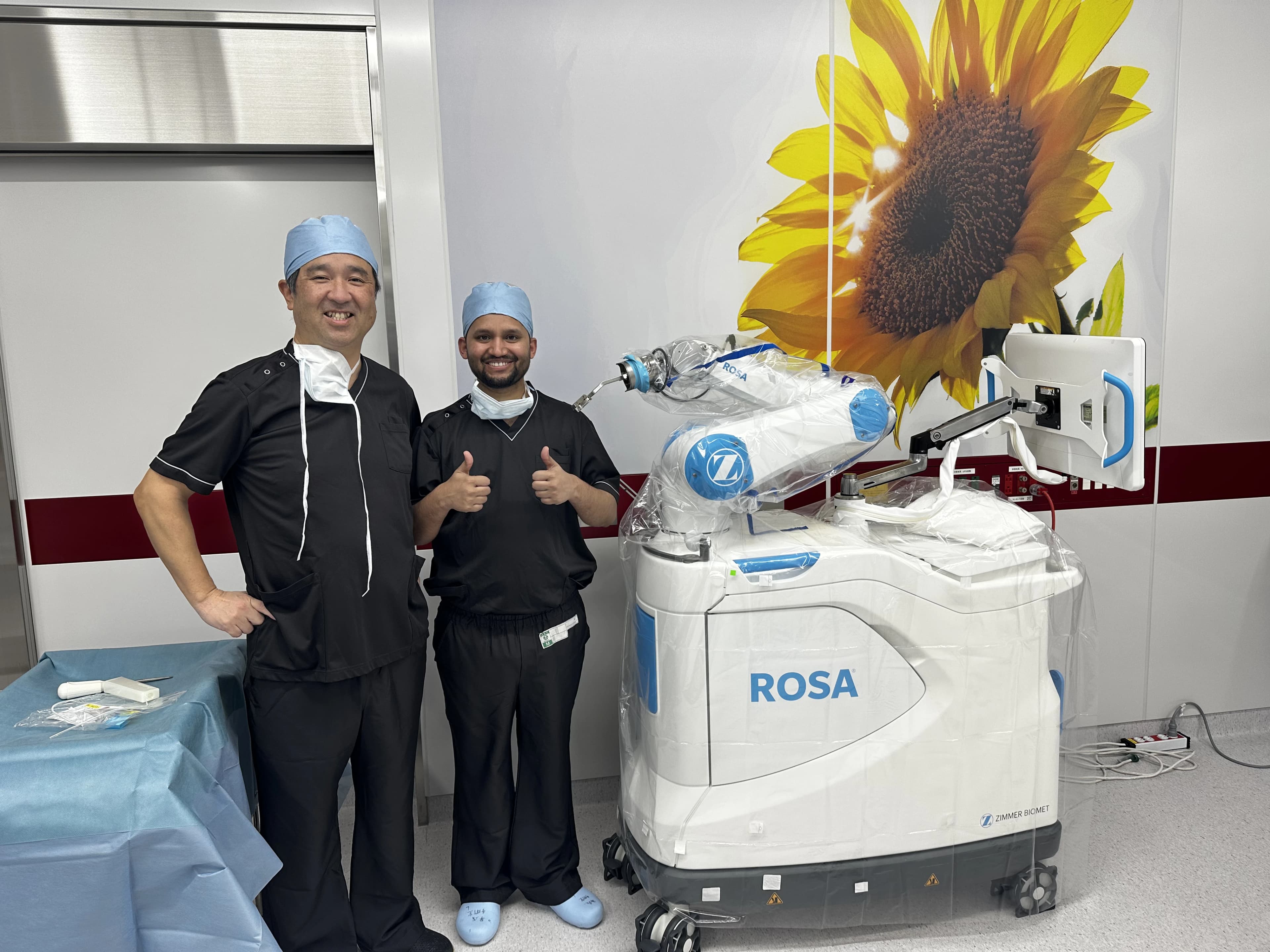
Intraoperative kinematics & 3D navigation alongside Japanese surgical faculty.
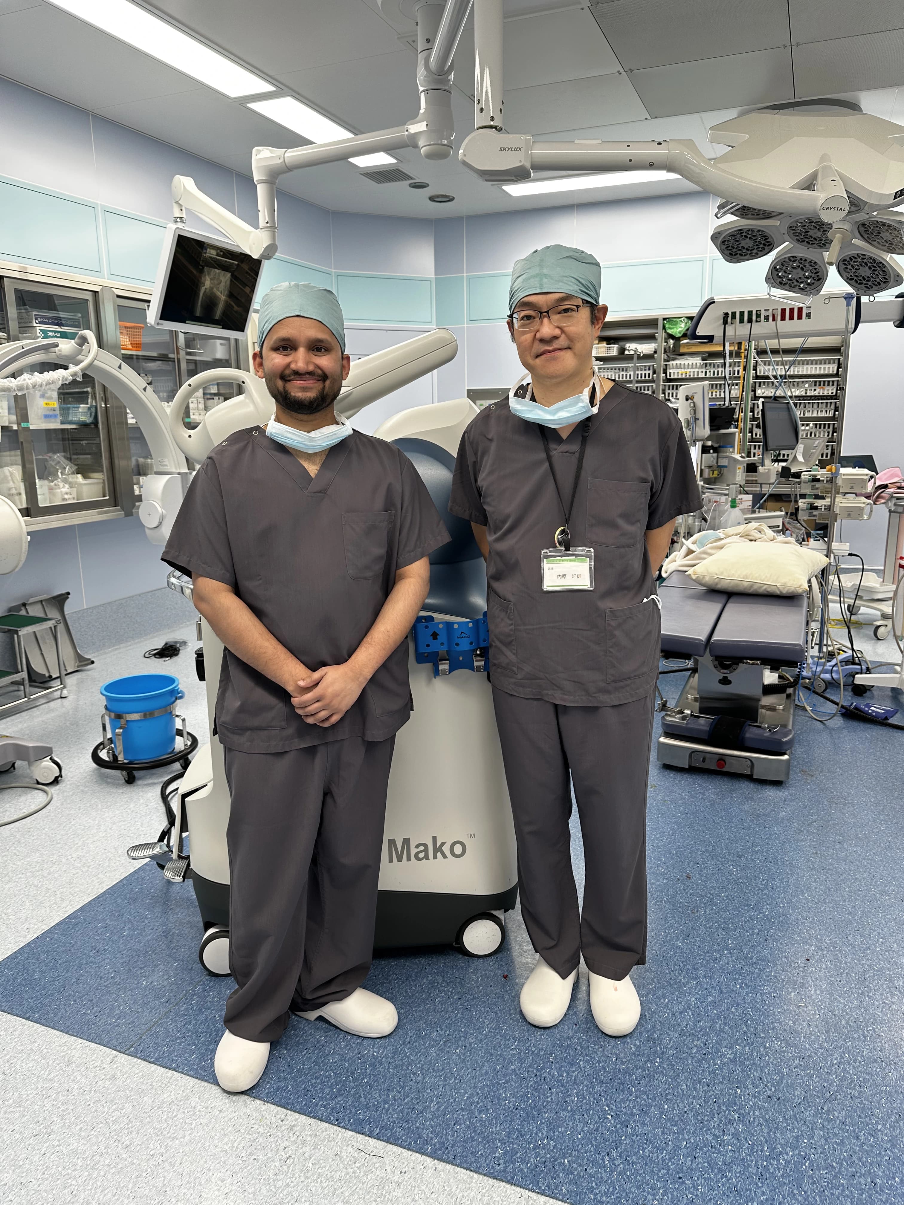
International fellowship-level precision applied to patient care in Patna.
Frequently Asked Questions on Patella Instability & MPFL
When performed anatomically with correct isometric femoral positioning, MPFL reconstruction has an outstanding success rate (>95%) with near-zero recurrence rates.