
Understanding Cartilage Defect Restoration & Meniscus Preservation
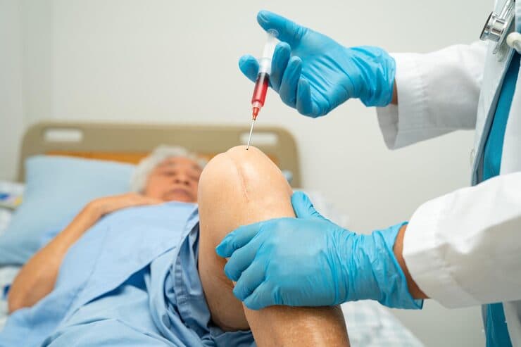
Articular cartilage is the ultra-smooth, low-friction lining that covers the ends of bones within joints. Because cartilage lacks direct blood vessels, focal injuries from sports impacts, twisting trauma, or osteochondritis dissecans (OCD) cannot heal spontaneously. Similarly, tears in the fibrocartilaginous meniscus reduce shock absorption, increasing stress on underlying bone by up to 300%. Modern joint preservation focuses on biological repair—using advanced suture techniques to save the meniscus and regenerative scaffolds or osteochondral transfers to resurface cartilage defects.
The Biological Joint-Sparing Philosophy
By correcting mechanical weight-bearing malalignment through precision osteotomy and cartilage preservation, the load is shifted off damaged cartilage onto healthy bone—preserving your natural joint for years to come.
When Should You Consider Consultation for Cartilage & Meniscus Repair?
Specialist intervention is considered when joint pain or functional limitation significantly disrupts your daily life and conservative measures have been exhausted. Check if you experience any of the following:
Persistent, localized knee pain following a sporting event, jump, or twisting injury.
Mechanical catching, clicking, or locking sensations when flexing or extending the knee.
Joint line tenderness and recurrent swelling after running, jumping, or prolonged standing.
Diagnosis of a focal cartilage defect (OCD lesion) or meniscus tear on an MRI scan.
Young or active patients seeking alternatives to premature joint replacement surgery.
Persistent symptoms after prior partial meniscectomy seeking joint-sparing solutions.
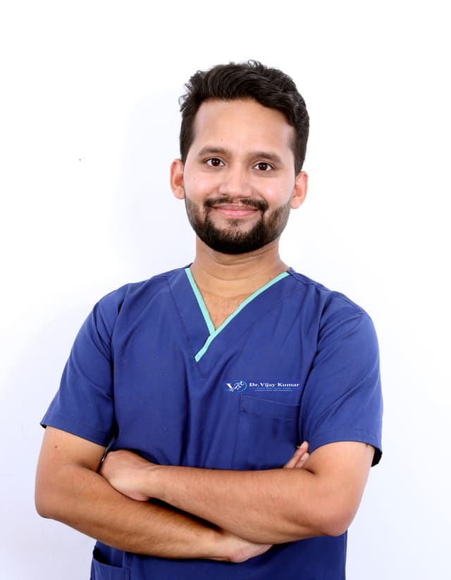
Dr. Vijay Kumar
Verified SpecialistSenior Consultant Orthopaedic, Robotic Joint Replacement & Complex Articular Trauma Surgeon
“My surgical philosophy centers on biological preservation, anatomical restoration, and rapid functional recovery. By tailoring the intervention to each patient's unique anatomy and lifestyle, we achieve lasting pain relief and active mobility.”
The 4-Stage Precision Clinical Workflow
Full-Leg Weight-Bearing Alignment Analysis
High-definition 4K arthroscopic assessment of the defect size, depth, and cartilage shoulder stability.
Precision Biplanar Osteotomy Planning
Preparation of the defect base down to healthy subchondral bone with micro-drilling to stimulate cellular repair.
Rigid Locking Plate Stabilization
Application of biological chondral scaffolds or transfer of cylindrical osteochondral autografts from low-weight-bearing zones.
Protected Unloading & Gradual Rehabilitation
High-tensile meniscal suture anchor repair securing torn fragments back to the vascular peripheral zone or bony root.
Stage 05: Joint Preservation Stage
Post-operative protocol combining continuous passive motion (CPM) with protected weight-bearing for optimal tissue integration.
Treatment Modalities Offered
Dr. Vijay Kumar practices conservative joint-sparing protocols first, proceeding to advanced surgical intervention only when indicated:
Non-Surgical & Joint Sparing
- Unloader knee bracing to shift weight off the damaged cartilage compartment
- Targeted neuromuscular physiotherapy to enhance joint stability and load absorption
- Orthobiologic injections (Platelet-Rich Plasma / PRP) to modulate joint inflammation
- Low-impact aerobic conditioning (cycling, swimming) to maintain joint nutrition
Specialised Surgical Procedures
- Arthroscopic Meniscus Suture Repair (All-Inside, Inside-Out, and Outside-In techniques)
- Meniscal Root Repair and Ramp Lesion Fixation
- Microfracture and Bone Marrow Stimulation with Collagen Matrix Scaffolding
- Osteochondral Autograft Transfer System (OATS / Mosaicplasty)
- Realignment Osteotomy (HTO / DFO) to de-load the repaired cartilage surface
Recovery Timeline & Mobility Milestones
Weeks 1–4: Continuous passive motion (CPM) to nourish new cartilage cells; protected touch-down weight bearing.
Weeks 6–8: Progression to full weight bearing, stationary cycling, closed-chain muscular activation.
Months 3–5: Progressive strengthening, low-impact elliptical and swimming workouts.
Months 6–9: High-level athletic conditioning, plyometrics, and clearance for return to running and sports.
International Fellowship & Surgical Suite Records
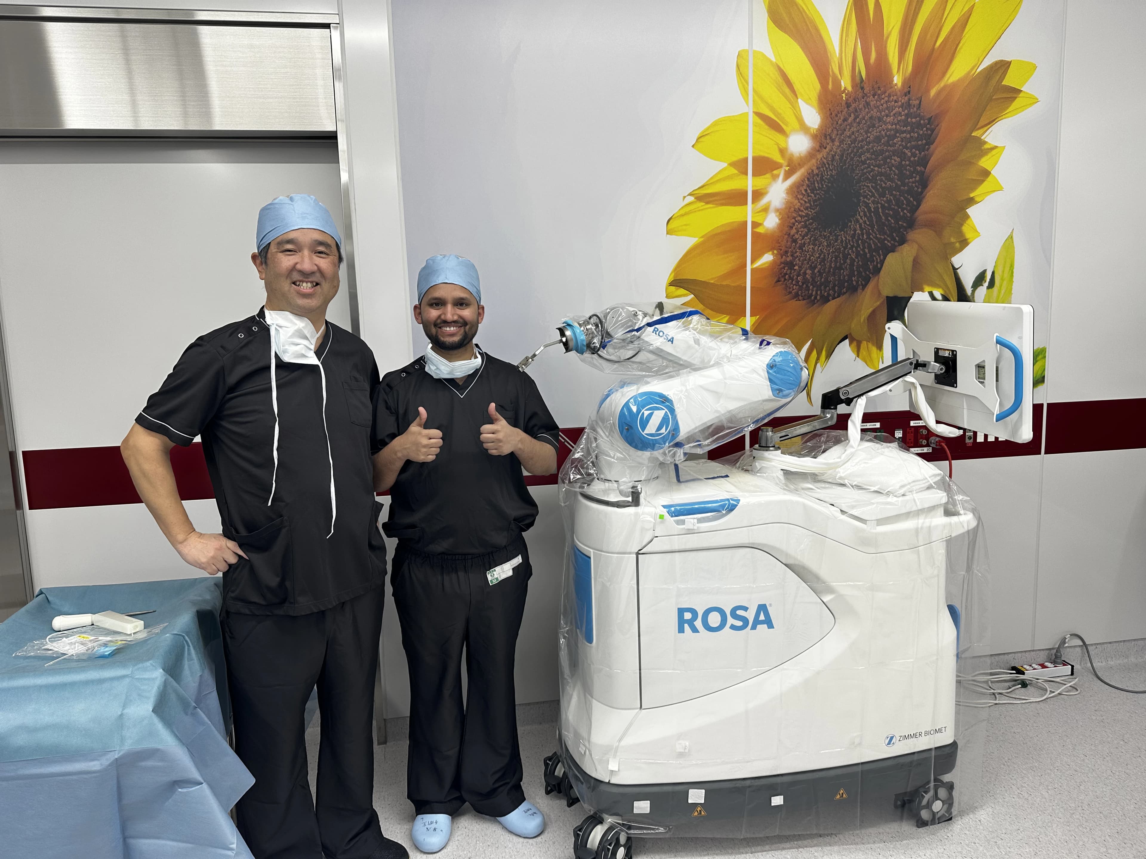
Intraoperative kinematics & 3D navigation alongside Japanese surgical faculty.
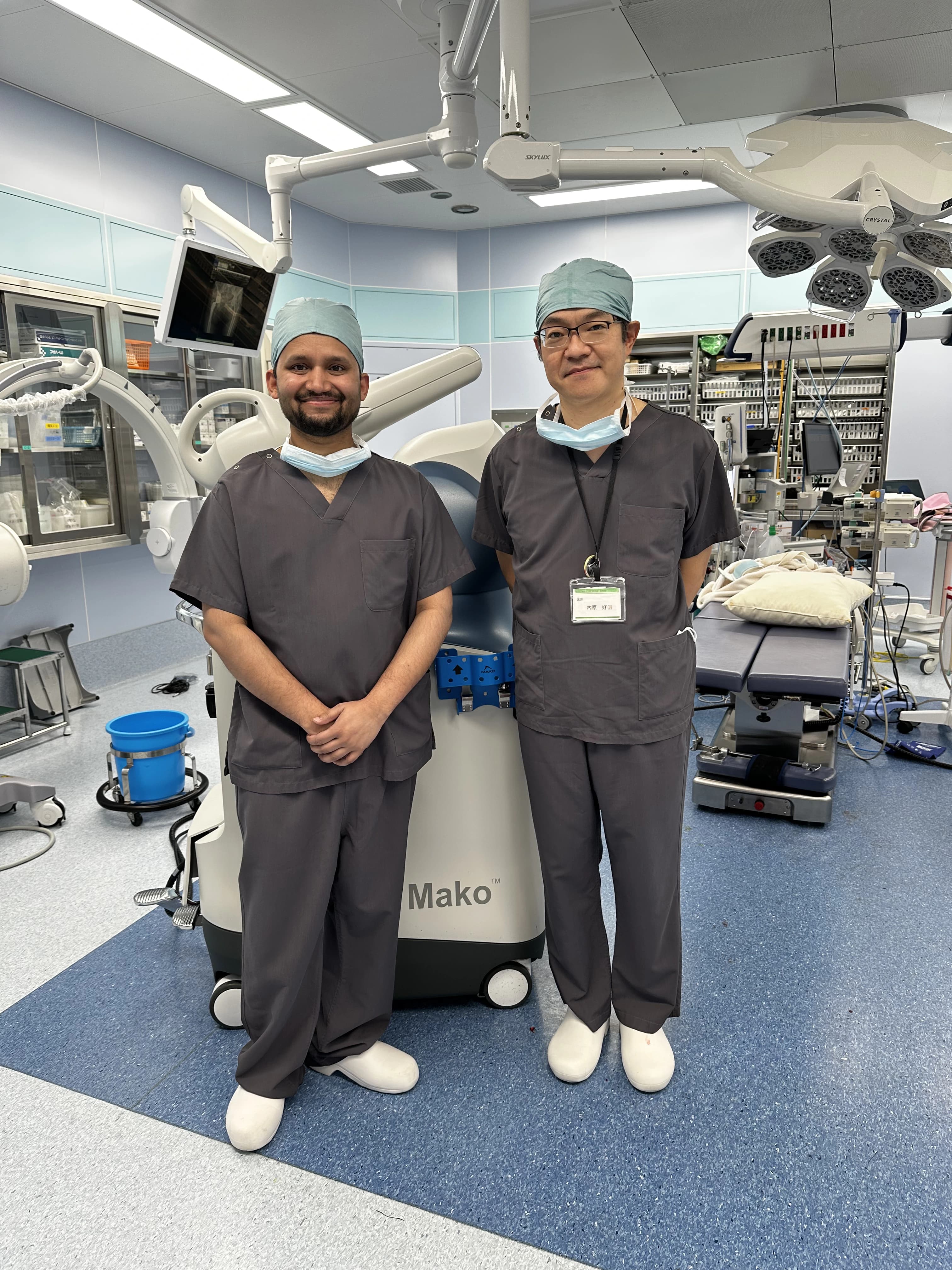
International fellowship-level precision applied to patient care in Patna.
Frequently Asked Questions on Cartilage & Meniscus Repair
The meniscus acts as the natural shock absorber and stabilizer of the knee. Removing it increases contact stress between femur and tibia by 200% to 300%, leading directly to rapid cartilage wear and early arthritis. Repairing the meniscus preserves natural joint biomechanics for life.