
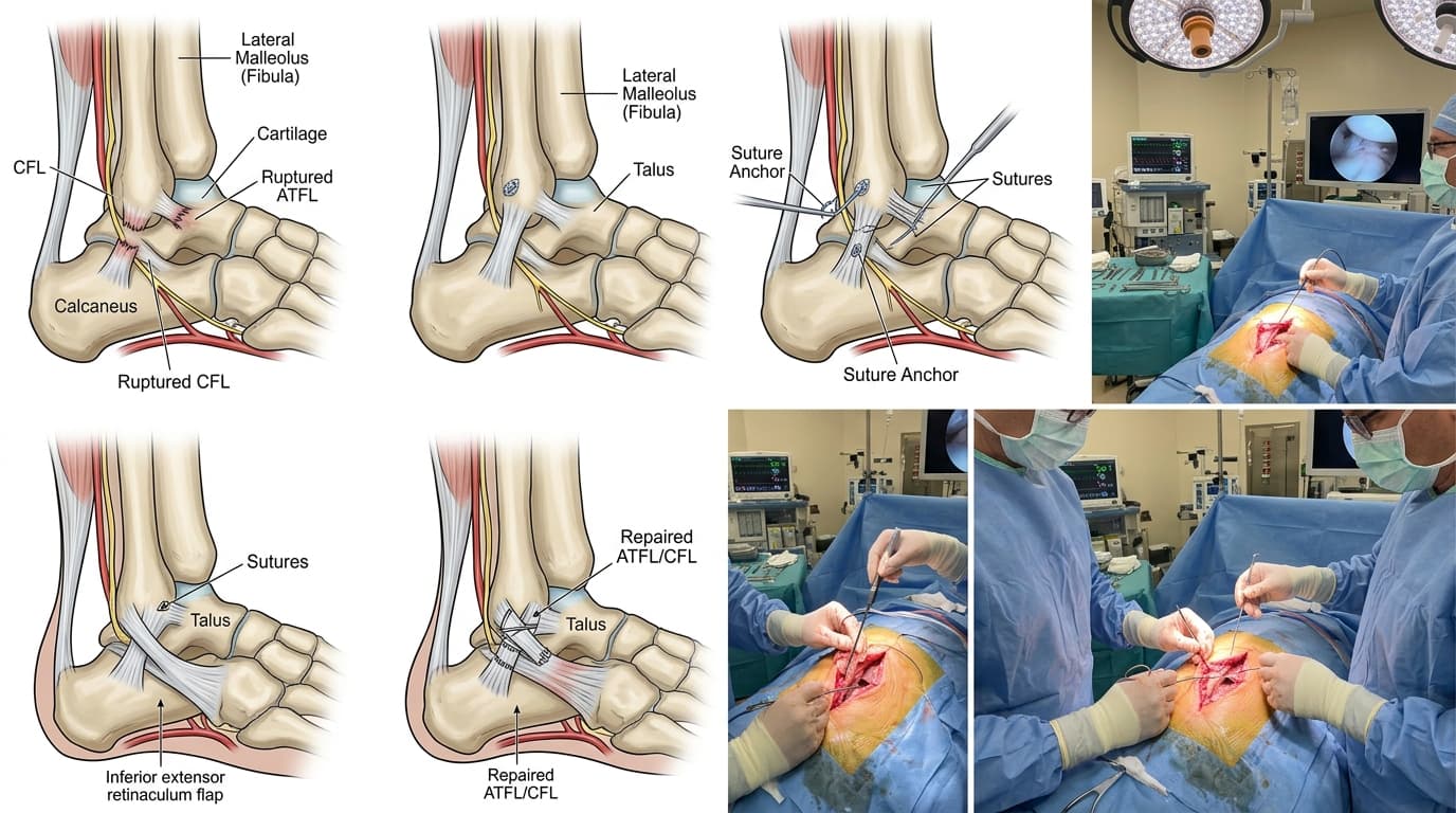

Understanding Ankle Ligament Reconstruction (ATFL / CFL) & Sprains
Ankle sprains are among the most common athletic and daily injuries, tearing the Anterior Talofibular Ligament (ATFL) and Calcaneofibular Ligament (CFL) on the outer ankle. While most acute sprains heal with conservative therapy, up to 20–30% develop Chronic Ankle Instability (CAI)—where stretched or ruptured ligaments fail to hold the ankle stable. Patients suffer recurrent twisting episodes, chronic swelling, and anxiety on uneven ground. The Modified Brostrom-Gould procedure with Internal Brace suture tape reconstructs the ligaments anatomically, restoring rock-solid stability.
The Minimally Invasive Arthroscopy Advantage
Advanced 4K keyhole arthroscopy allows complete visualization and anatomical repair through pencil-thin incisions, eliminating extensive soft-tissue cutting, preserving natural joint biomechanics, and drastically accelerating recovery.
When Should You Consider Consultation for Ankle Ligament Surgery?
Specialist intervention is considered when joint pain or functional limitation significantly disrupts your daily life and conservative measures have been exhausted. Check if you experience any of the following:
Ankle that frequently twists, rolls over, or 'gives way' during walking, running, or stepping off curbs.
Persistent aching pain and swelling on the outer ankle months after a sprain.
Apprehension and lack of confidence when walking on grass, cobblestones, or uneven ground.
MRI confirmation of complete ATFL/CFL tear or attenuation.
Athlete or active individual unable to return to basketball, football, running, or tennis due to ankle instability.

Dr. Vijay Kumar
Verified SpecialistSenior Consultant Orthopaedic, Robotic Joint Replacement & Complex Articular Trauma Surgeon
“My surgical philosophy centers on biological preservation, anatomical restoration, and rapid functional recovery. By tailoring the intervention to each patient's unique anatomy and lifestyle, we achieve lasting pain relief and active mobility.”
The 4-Stage Precision Clinical Workflow
Pre-Operative MRI & Diagnostic Mapping
4K diagnostic ankle arthroscopy evaluating cartilage integrity and clearing anterolateral soft-tissue impingement.
High-Definition 4K Arthroscopic Access
Minimally invasive 3-cm incision over the lateral malleolus.
Precision Anatomical Repair / Reconstruction
Identification and anatomical mobilization of the torn ATFL and CFL ligaments.
Bio-Fixation & Sports Rehab Roadmap
Re-anchoring ligaments firmly to the fibula using bio-composite suture anchors and reinforcing with inferior extensor retinaculum.
Stage 05: Arthroscopic Execution
Application of high-strength Internal Brace suture tape bridging the fibula to talus for immediate mechanical protection.
Treatment Modalities Offered
Dr. Vijay Kumar practices conservative joint-sparing protocols first, proceeding to advanced surgical intervention only when indicated:
Non-Surgical & Joint Sparing
- Neuromuscular peroneal muscle strengthening and wobble-board balance therapy
- Semi-rigid functional lace-up ankle braces during athletic activity
- Custom orthotic insoles with lateral heel wedges
- Activity modification avoiding high-risk unbraced sports
Specialised Surgical Procedures
- Modified Brostrom-Gould Anatomical Ligament Repair
- Internal Brace Suture Tape Augmentation (allowing early accelerated mobilization)
- All-Arthroscopic Brostrom Repair with bio-anchors
- Anatomical Tendon Autograft Reconstruction for long-standing neglected laxity
Recovery Timeline & Mobility Milestones
Weeks 1–2: Protected weight-bearing in walking boot, gentle active ankle pumping exercises.
Weeks 3–4: Weaning off boot into supportive athletic shoe, stationary cycling, balance board training.
Weeks 6–8: Agile lateral movement drills, jogging, resistance band peroneal strengthening.
Weeks 10–12: Full return to competitive athletics, tennis, football, and running without brace.
International Fellowship & Surgical Suite Records
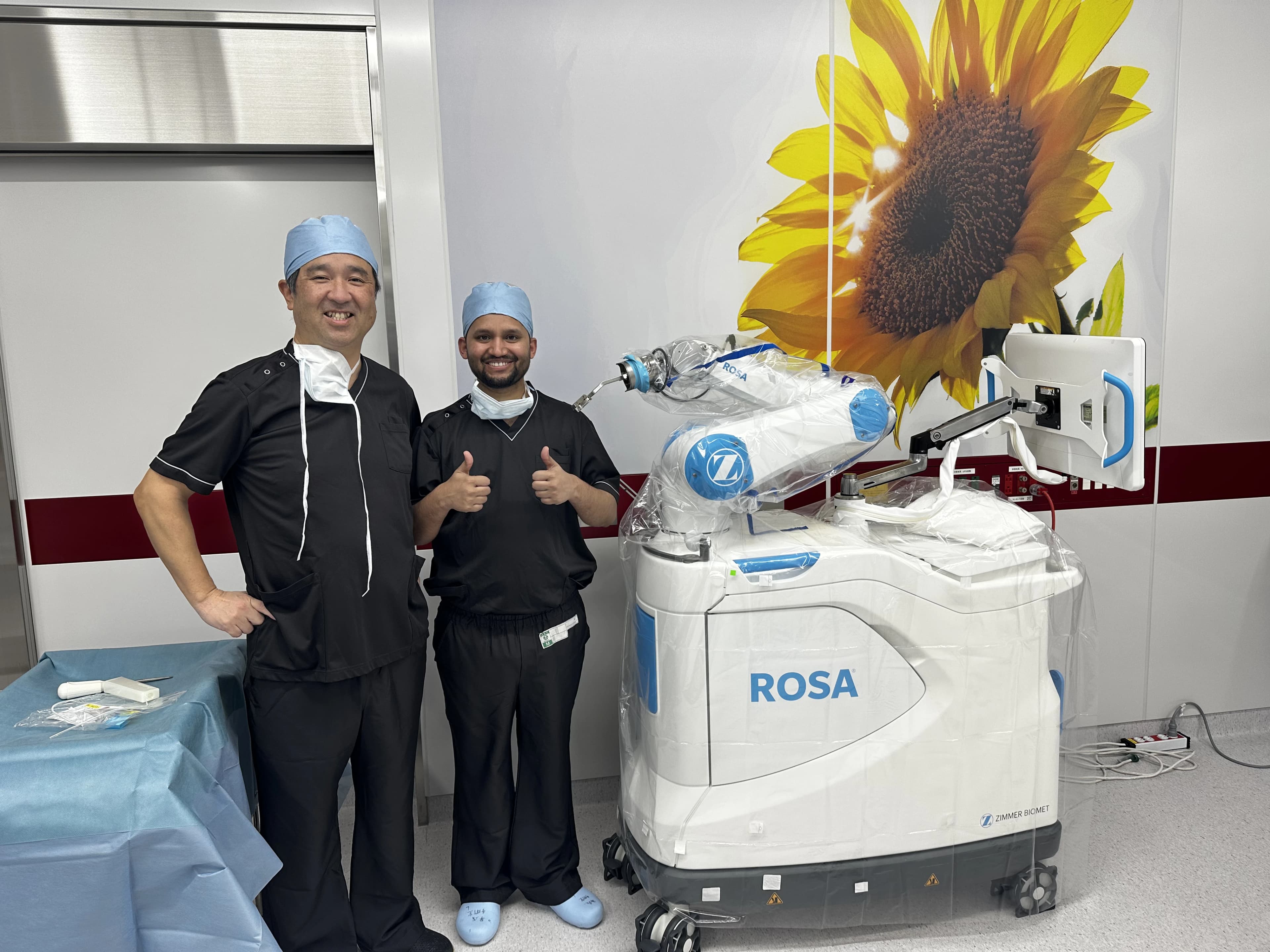
Intraoperative kinematics & 3D navigation alongside Japanese surgical faculty.
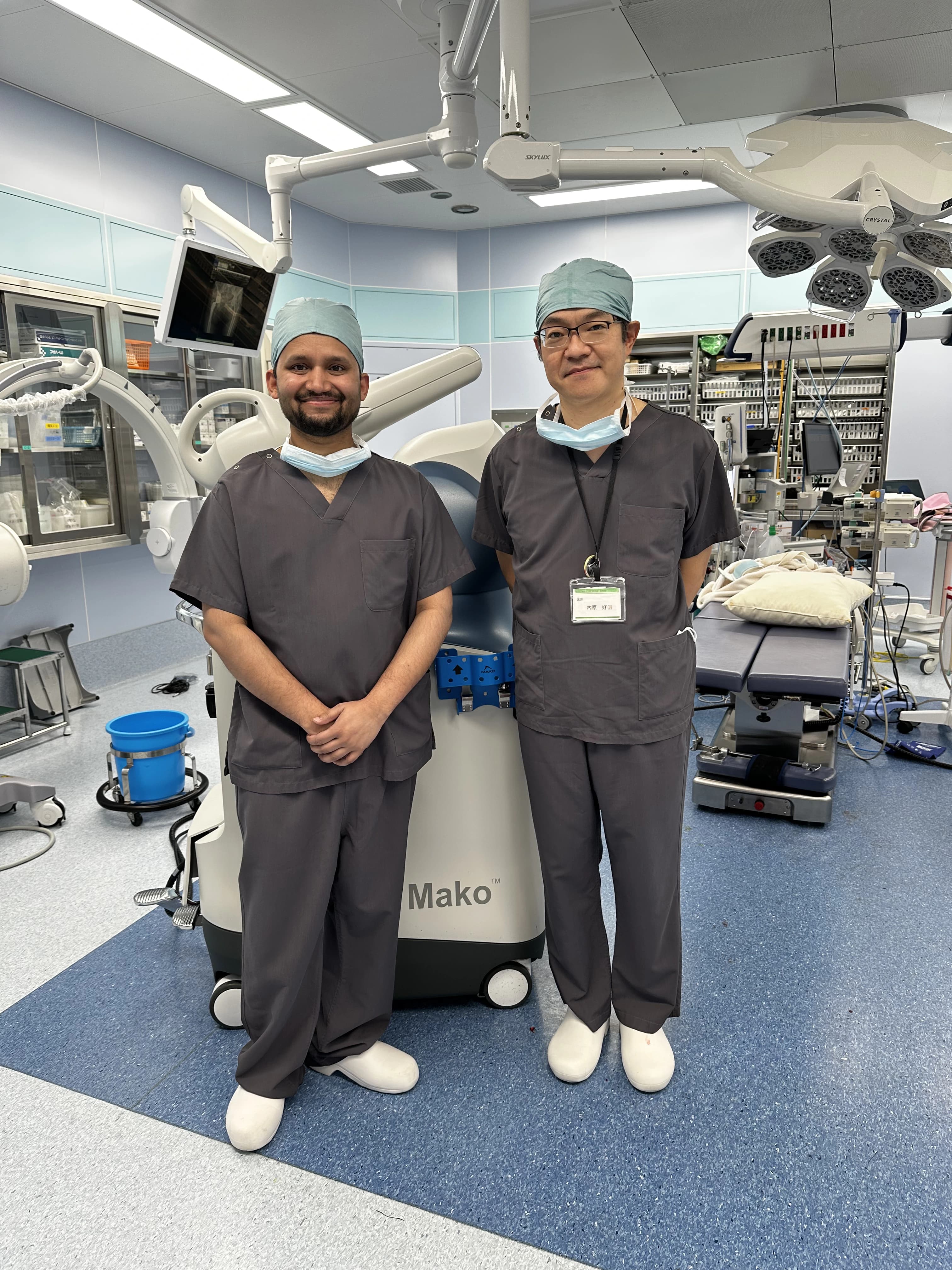
International fellowship-level precision applied to patient care in Patna.
Frequently Asked Questions on Ankle Ligament Surgery
The Internal Brace is a high-strength braided suture tape placed over the repaired ligament that acts like a secondary seatbelt, protecting the healing tissue and allowing patients to walk weeks earlier.